MOHS Surgery in Mobile, Eastern Shore, Daphne AL.
 What to expect with Mohs Surgery
Mohs Micrographic Surgery (MMS), named for its originator Dr. Frederic Mohs, is a highly specialized surgical technique for treating skin cancer where the doctor performs the role of both surgeon and pathologist. The technique was developed in the 1930s and is currently used around the world. Mohs surgery allows for the immediate microscopic examination of the removed cancerous tissue which eliminates days of waiting for a pathology report from an outside lab.
During MMS, the entire border between the cancerous skin and your healthy skin is evaluated, giving you the highest cure rate possible. This is what “100% surgical margin evaluation” means. The MMS technique works well because cancer grows and spreads in a haphazard fashion, making it hard to differentiate from healthy skin without precise microscopic evaluation.
MOHS Micrographic Surgey Benefits:
We perform Mohs Micrographic Surgery because of its benefits which include:
- Precise removal of cancerous tissue
- Maximum preservation of uninvolved, healthy tissue
- The highest cure rate of all skin cancer treatments
- Performed by a fellowship trained Mohs surgeon, Mohs Micrographic Surgery is the “gold-standard” for the treatment of skin cancer
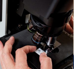 First, the affected area is numbed. Then, a thin layer of tissue is removed from the surrounding skin and base. The removed tissue is mapped and sectioned by your surgeon. The tissue is taken to our in-house lab where the deep and peripheral margins (borders) of each section are thinly sliced and mounted on microscope slides. Next, your surgeon examines the tissue under the microscope. If additional cancer is found at the margin, your surgeon notates this on the map, and the process continues until the surgical margins are completely clear.
There are two important distinctions between Mohs surgery and other techniques.1. 100% surgical margin evaluation 2. The doctor performs the role of both surgeon and pathologist
Below are 4 steps in the Mohs process.
1. Surgical removal of the cancerous tissue
- The surgeon identifies the cancerous lesion
- Your affected area is anesthetized or numbed
- The visible cancer and a thin margin of additional tissue are removed
2. Mapping of the tissue and surgical site, and processing the tissue
- The surgeon maps, or creates a diagram, of the area
- The tissue is processed and slides are prepared for microscopic analysis
3. In-house interpretation of microscope slides
- The Mohs Surgeon examines the tissue with a microscope, taking note of any remaining cancerous cells and precisely recording this on the map
- The patient returns to the operating room in order to remove the cancerous tissue
- The map directs the surgeon to the exact location of the remaining cancer, sparing precious normal skin
- Mapping, slide preparation, and microscopic analysis are performed again
- This process will continue until the surgeon determines that the margins are clear
4. In-house repair of the surgical defect
- Once your cancer has been completely removed, your surgeon will determine the best method for repairing your wound or “defect”
- In most cases, your surgeon will perform the reconstructive procedure on the same day that your cancer has been removed
During MMS, the entire border between the cancerous skin and your healthy skin is evaluated, giving you the highest cure rate possible. This is what “100% surgical margin evaluation” means. The MMS technique works well because cancer grows and spreads in a haphazard fashion, making it hard to differentiate from healthy skin without precise microscopic evaluation.
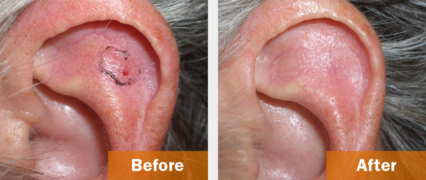
We believe in providing our patients the best reconstructive service possible. This is to create as little change possible both cosmetically and functionally to sensitive areas, such as the lips, nose, or ears. We are proud of our results. On this page, you can see actual patient before and after photographs from Dr. Henghold.
Before & After Surgery Patient Photos
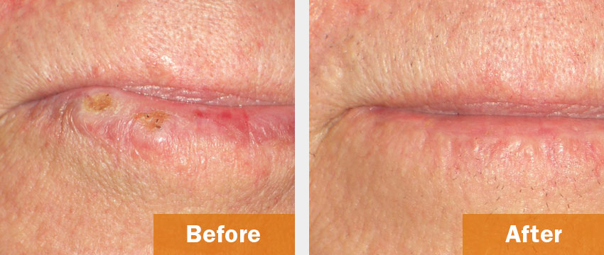
This patient was afflicted with Squamous Cell Carcinoma on their lip. The after picture showcases the tissue saving abilities of Mohs Micrographic surgery and the skill of our reconstructive surgeons in preserving your face.
*Pictures have not been altered.
Mohs Surgery Instructions – Download
FAX: Alabama-251-631-3572 or 251-621-7209
Florida is 850-266-6301